
Benign paroxysmal positional vertigo (BPPV)
BPPV is one of the most common causes of dizziness. This is a good example of paroxysmal dysfunction of the inner ear.
1. Posterior canal BPPV
The patient complains of intense rotational vertigo associated with movements of the head with three characteristics:
1. it is only triggered when the position of the head is moved,
2. it is very violent,
3. it lasts for less than one minute,
4. it can cause nausea and vomiting.
The head positions that trigger vertigo are most often those where the head is extended or when the patient goes to bed at night or goes from lying to sitting in the morning. The vertigo recurs whenever the patient’s head returns to the same position but becomes less intense, reflecting its fatigability. Physician-patient discussion will be immediately helpful but only the clinical examination will confirm diagnosis.
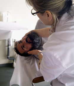
The examination must be performed using VNG and will involve positioning manoeuvers. In practice, the patient will sit across the bed and switches quickly from sitting to lying on their side (on the side that triggers dizziness), the head unsupported and turned 45°. After a few seconds, nystagmus and rotational vertigo are triggered. Nystagmus and dizziness stop after around ten seconds if the head is kept in the same position. Sitting up again triggers another nystagmus, this time in the opposite direction, and also accompanied by a rotational sensation. The repetition of these manoeuvres leads to a gradual decrease in the nystagmus and vertigo. Vertigo and nystagmus are therefore fatigable. Diagnosis of this vertigo is therefore based on both physician-patient discussion and clinical examination.
Physiopathology
Physiopathogenesis is now well established and incriminates a condition in the posterior semicircular canal, itself induced by an impairment of the statolith in the utricle. This lesion is characterized by the otoconia of the statolith in the utricle becoming detached, and then being deposited on the most inclined part of the labyrinthine cavity, i.e. on the ampulla of the posterior canal. This detachment can be caused by a trauma, virus, infection or may be degenerative.
Treatment
The treatment depends on the stage at which the patient is seen. If the patient sees the physician during episodes or a relapse, treatment is based on physiotherapy.
Liberatory manoeuvre
The liberatory manoeuvre consists in moving the patient’s head vigorously to displace the cupulo- or cana-lithiasis. This manoeuvre often cures positional vertigo. It can be repeated during another session if the positional vertigo is still present. If the positional vertigo is refractory to two or three correct manoeuvres, the practitioner should revise the diagnosis. These manoeuvres should never be repeated frequently. However, it is not uncommon to see patients who have undergone over 10 manoeuvres complaining of a permanent feeling of inebriation.
Si le patient est vu à distance de la crise, un bilan de l’équilibre doit être fait sur l’équitest. Ce dernier permettra de quantifier les troubles de l’équilibre et de prescrire des séances de rééducation et une thérapie médicale efficace.
2. Positional vertigo of the horizontal canal
This type of positional vertigo is much rarer. It most often occurs not when the patient lies down or stands up but when they are already lying down and turn over to one side or the other. Clinically, diagnosis is based on several types of argument:
1. it is not triggered by lying on the side as for the posterior canal vertigo, but occurs when patients sit with their head bent forward, placing the horizontal canals in a vertical plane;
2. the nystagmus induced is horizontal and not torsional. In this position, the direction of the rapid phase of nystagmus indicates the affected side;
3. it is rarely fatigable;
4. its onset latency is shorter (less than 5 s) and its duration is longer (20-60 s).
Treatment is based on a slightly different liberatory manoeuvre: the “barbecue” manoeuvre.
In conclusion
All types of positional vertigo should be treated to prevent anxiety or reactive depression associated with poor quality of life. A full otoneurological assessment (see otoneurological exploration) must be performed at a distance from the episode to try to ascertain the cause of the positional vertigo and prevent a recurrence. There should be no room for doubt, and the patient should not have to settle for preventive measures such as lying down slowly or not moving their head, etc.