Functional anatomy of the inner ear
HISTORICAL REVIEW
Before describing the anatomy of the inner ear, we will briefly review the relatively recent discovery (1914) of the "vestibule".
- 535 BC: The vestibule was discovered very late. It was considered by Empedocles (535-475 BC) to be a hearing organ.
- 1540. In the 16th century, Gabriel Fallope gave the first complete anatomical description of the inner ear, distinguishing its two cavities: the labyrinth and the cochlea.
- 1800. In the 19th century, Pierre-Jean Marie Flourens mentioned that the labyrinth is the peripheral organ in which the moderating forces of movement reside.
- 1861. The vestibular labyrinth did not appear in the clinical setting until 1861, following the famous report by Prosper Meniere, which described the first vertiginous syndrome related to the inner ear (Meniere's disease).
- 1875. The geometry of the semi-circular canals, in particular their respective orientation, as well as their functioning as a pair, was described by the German physicist Ernst Mach in 1875.
- 1914. In 1914, Robert Barany won the Nobel Prize in Physiology or Medicine “for his work on the physiology and pathology of the vestibular apparatus”.
Description
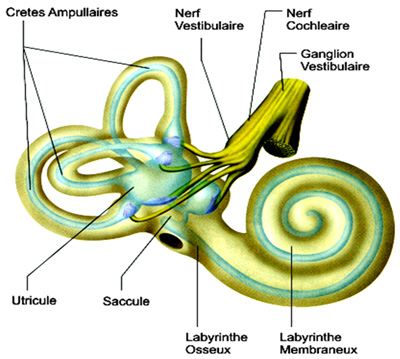
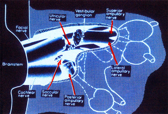
The peripheral vestibular system is included of five different types of sensors in each ear: three semi-circular canals (horizontal, and anterior and posterior vertical canals) and two otolithic organs (the utricle and the saccule), i.e. a total of ten receptors. The semicircular canals detect the amplitude of the angular rotation of the head in the three dimensions of space. The otolithic organs are sensitive to vertical linear acceleration (the saccule) or horizontal linear acceleration (the utricle) of the head in space, and they detect tilting of the head relative to gravity.
Inner hear
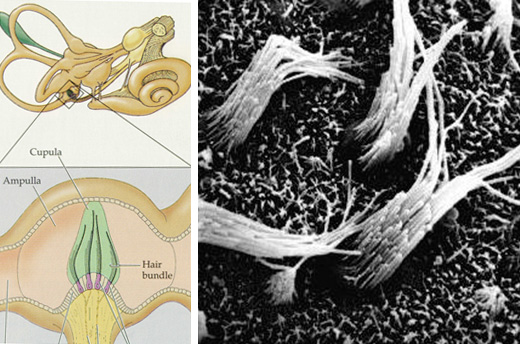
Canal hair cells
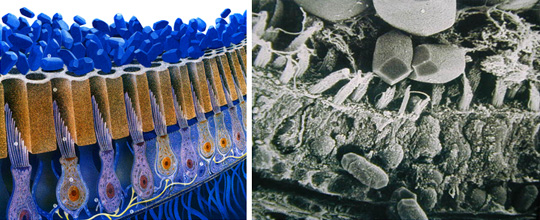
Sensory cells are hair cells whose cilia bathe in the endolymph. Otolithic sensory cells have the characteristic of being covered by a tectorial membrane embedded with small crystals of calcium carbonate or otoconia.
The sensory cells send information to the central nervous system via the vestibular nerve, which has two parts:
• the superior vestibular nerve comprising the nerves of the horizontal and anterior canals and utricular nerve.
• the inferior vestibular nerve comprising the saccular nerve and posterior canal nerve.
The vestibular nerve
Vestibular nuclei
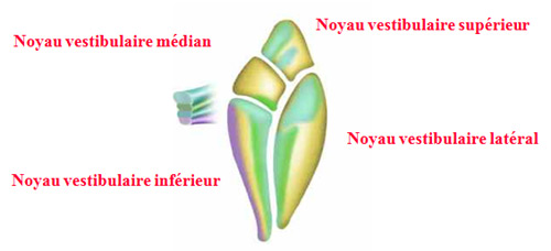
The two branches of the vestibular nerve are distributed to the vestibular nuclei, located in the brain stem either side of the fourth ventricle.
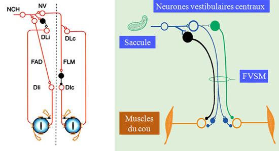
The central vestibular neurons are then distributed to the oculomotor nuclei to steady the gaze or to the spine to stabilize posture.
Vestibulo-ocular and vestibulo-spinal pathways
In addition to vestibular inputs, these nuclei receive visual and proprioceptive inputs. Vestibular nuclei are therefore not merely there to relay information from the inner ear, but are veritable sensory motor integration centres. The central vestibular neurons are then distributed to the oculomotor nuclei to steady the gaze or to the spine to stabilize posture.
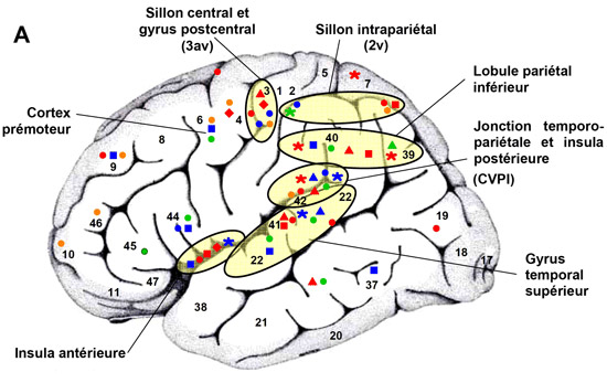
Projections corticales vestibulaires
The vestibular nuclei are also distributed to the cortex, which plays a part in the perception of movement.